Northeastern Researchers Develop Breakthrough fMRI Technique
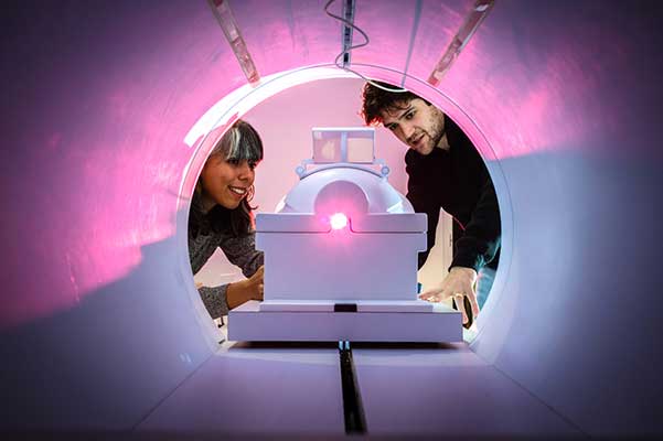
COS/BioE Assistant Professor Stephanie Noble developed a functional MRI breakthrough that enhances the precision and speed of brain imaging, opening new possibilities for mapping neurological activity and understanding cognitive functions.
This article originally appeared on Northeastern Global News. It was published by Noah Lloyd. Main photo: Stephanie Noble, left, with co-author Raimundo Rodriguez, conducted functional MRI research that “caricatures” the wiring diagrams of the brain. Photo by Alyssa Stone/Northeastern University
How a ‘caricature’ of a brain scan could lead to better treatment outcomes
New research removes task-based information from resting-state MRI scans, leading to more individual diagnoses and better predictions.
New research suggests that simplifying how brain imaging data are analyzed could make functional MRI scans far better at predicting individual health outcomes, a counterargument to the “just in case” MRI.
According to the Journal of Magnetic Resonance Imaging, elective, preventative MRI scans are often rife with false positives, which can be costly in both a financial and emotional sense.
In a typical functional magnetic resonance imaging study, or fMRI, you’ll be asked to lay down in an MRI scanner while either remaining at rest or performing a specific task. A new paper from Stephanie Noble, assistant professor of psychology and bioengineering at Northeastern University, and visiting Ph.D. student Raimundo Rodriguez, identifies signal information characteristic of performing tasks. By removing this information from resting-state models, their research finds that resting-state brain maps produced by fMRI highlight individual differences and become better suited to predicting information about the subject.
With further research, these simplified maps could mean earlier predictions of mental disorders like schizophrenia and an increased understanding of which treatments would best suit particular patients.
Connecting over connectomes
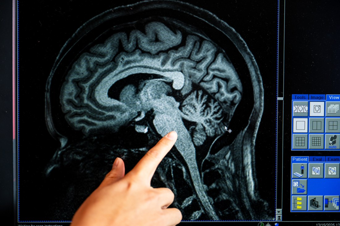
Whenever you think, areas of your brain flush with blood. MRI machines measure the blood flow, which Noble says is like a movie of brain activity, to build interlinking maps of the brain called “connectomes.”
These maps contain huge amounts of data and are “very complex, very high-dimensional,” Rodriguez says. The maps are so complex and information-rich, in fact, that researchers are still discovering ways to use them.
Rodriguez says that functional MRI experiments are undertaken with subjects in one of two states: either resting or involved in a simple task of some kind, like pushing a button in response to stimuli or working memory exercises, he says.
 |
 |
 |
|
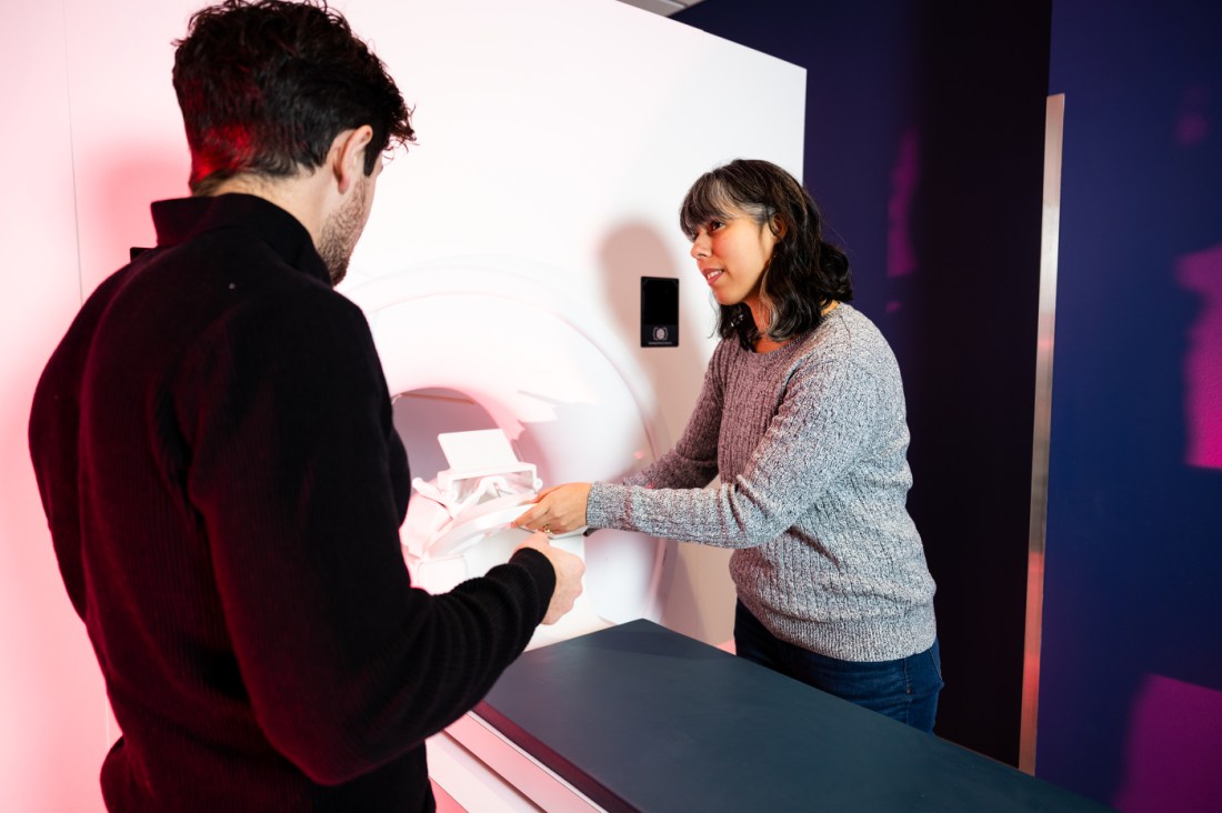
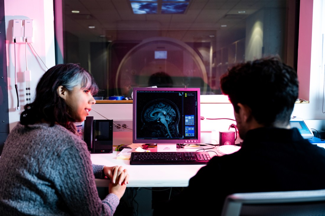
“If you can make people look really different from each other, that has the potential to increase your ability to find relationships between people and outcomes you care about,” Noble, bottom left, says. Rodriguez says that, by highlighting specifics, caricatured brain maps can be used to identify patterns previously obscured by the more information-laden connectomes. Photos by Alyssa Stone/Northeastern University
The task-based scans, especially, show the human brain in action. But even at rest, the brain is active and contains information that looks similar to task-based signals.
Previous work, he continues, noted that task-oriented connectomes, across multiple subjects, contain the same patterns of information. But what at first appears to be purely task-based data also appears, quite prominently, in resting states as well.
Read full story at Northeastern Global News