An eye toward better treatment
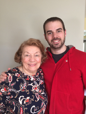
Every two months, Northeastern bioengineering graduate student David Walsh’s 91-year-old grandmother goes to the doctor to receive a drug injection into her eyes. She has wet age-related macular degeneration. There is no cure, only this invasive, recurring treatment.
“She worries a lot because she goes in, they inject her, and she leaves, and since the effect of the drugs is so gradual, she doesn’t know if it’s working or not,” said Walsh. “She worries that maybe she got too much drug, not enough drug, or if the drug is doing anything at all for her condition.”
To solve this problem, Walsh is developing a device that will provide valuable feedback to patients such as his grandmother and their clinicians.
As a member of associate professor of chemical engineering Shashi Murthy’s lab, Walsh helps design microfluidic devices that use a single drop of blood or other bodily fluid to diagnose a range of diseases. In work recently reported in the journal Lab On a Chip, Walsh and his colleagues have created a device that monitors the efficacy of treatments for two eye diseases: age-related macular degeneration and diabetic retinopathy.
“These are diseases where little blood vessels grow in the back of the eye and can pop your retina off or obscure your vision,” Walsh explained. This, he said, is due to increased levels of a molecular biomarker called vascular endothelial growth factor, or VEGF.
The standard treatments for these two eye diseases involve using drugs that bind to VEGF, thereby blocking its ability to interact with the cellular environment and causing blood vessel growth. These drugs are injected directly into the eye typically every four to six weeks, and often over the course of a lifetime. Walsh said the invasive nature of treatment and lack of personalized dosage raise patient risk and discomfort. What’s more, clinicians don’t usually test to confirm the efficacy of treatment.
That’s because the current diagnostic protocol—ELISA, or enzyme-linked immunosorbent assay—is extremely expensive and time consuming. Unless there’s no visible improvement after many injections, it doesn’t make sense to do an ELISA test, Walsh explained.
That’s where his device is different. Each time a patient undergoes a round of treatment, which includes a drug injection directly into the eye, a clinician can also take a very small sample of eye fluid—which contains a plethora of information—and test it in house in fewer than 20 minutes.
The device works like a light switch, where the on-button is only activated when VEGF is present. And the more VEGF, the brighter the light.
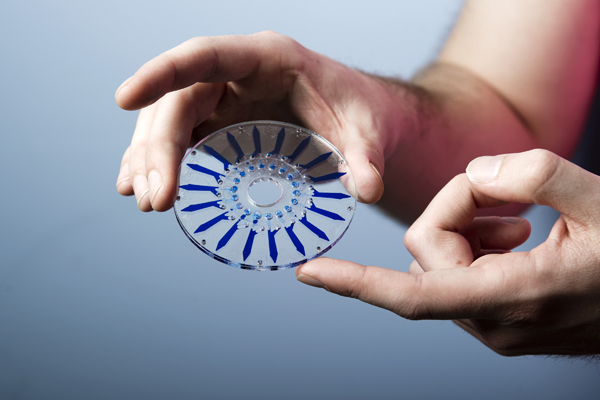
It’s made of two thin disks just a few inches wide, held together by double-sided sticky tape that’s been expertly etched into a specific design. That design creates an array of microscopically thin hollow channels into which Walsh inserts three reagents of varying density. The first is the densest and contains a photoactive substrate that glows in the presence of a particular enzyme. The second layer simply acts as a density gradient, while the third layer is the ocular fluid sample that Walsh wants to test. This layer also contains the enzyme as well as some tiny dense beads. Both of these are linked to an antibody specific to VEGF, which allows them to form a complex with the biomarker.
Spinning the whole device at very high speed allows these dense complexes to migrate to the layer containing the substrate where the enzyme causes it to ignite. But if there’s no VEGF in the sample, the enzyme stays in the least dense layer and no light is produced.
The team collaborated with clinicians at the Duke Eye Center in Durham, North Carolina, to obtain fluid samples with which to test the device. It passed with soaring colors, distinguishing not only between patients with age-related macular degeneration or diabetic retinopathy and patients with other diseases, but also between patients who have active and inactive forms of either of these two eye diseases.
The devices cost less than $1 and take minutes to assemble and run. With this platform, a clinician wouldn’t need a compelling reason to test whether treatment for these two eye diseases is effective—instead, she could look at a patient’s VEGF levels every time he came in for treatment. Blood vessel growth without VEGF would be a strong—and early—sign that something else was going on. What’s more, lack of reduction of VEGF over a few treatments would indicate a non-responder and allow the clinician to switch to another drug or treatment.
Information like this could not only help put Walsh’s grandmother—and patients like her—at ease. It could also help diagnose more serious conditions earlier.
Separately, Walsh also recently earned a NSF Graduate Research Opportunities Worldwide grant to perform related ocular diagnostic research at the KTH Royal Institute of Technology in Stockholm, Sweden. He will begin that work in September.